decoration
irys-header
Software

iRYS

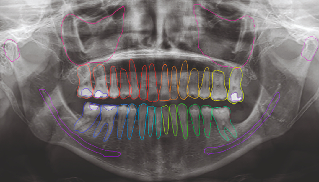
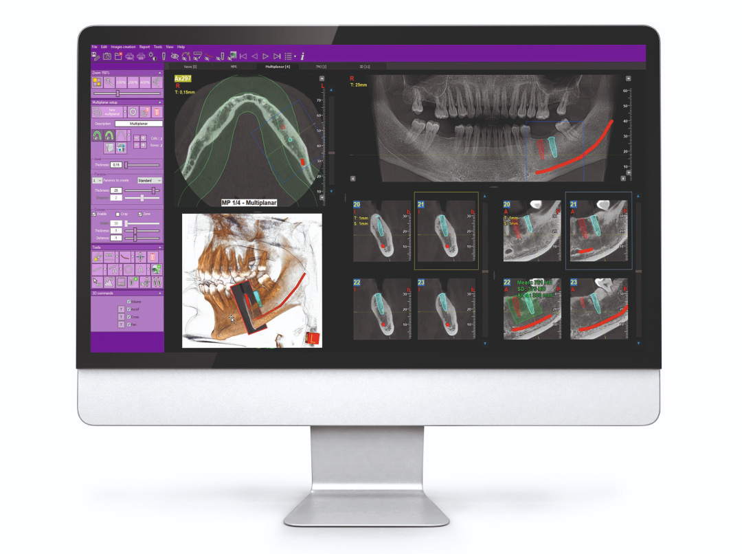
The best all-in-one software platform for 2D and 3D imaging. iRYS is data protection-certified and is IHE compliant with DICOM networks.

Characteristics

iRYS is a state-of-the-art tool equipped with a complete ecosystem of features to view, process and share scans directly from the dedicated workstation, from the dental practice computers or via the iRYS Viewer application available for iPAD.

Dose Book Tool

A feature designed to make iRYS software more efficient at gathering and managing information on acquired scans: the system allows automatic extraction and storage of data relating to the patient dose and other relevant information for each scan.
Users can also easily select information relating to the specific scan and filter the list of patients by category or date.
Lastly, the system lets you export data in .pdf or .csv format for quick and easy sharing.

MyRay has chosen to integrate Dentaldose to provide you with the most effective solution for managing your diagnostics exposure data.

Multi-Desktop 3D/2D

One software to manage and process 3D and 2D images: the iRYS Multi-Desktop system allows rapid browsing of the different 2D and 3D views, with realistic rendering and multiplanar analysis. Everything you need to carry out high-quality diagnoses and communicate with the patient quickly.

Preloaded implant libraries

iRYS streamlines the selection and positioning of implants chosen from those in its extended library. You can also modify options or add new ones in a few simple steps.

Diagnosis and planning

iRYS simplifies diagnosis and planning, with cutting-edge protocols and filters taking efficiency to the next level.
irys-3dsmart

3D SMART

The 3D SMART (Streak Metal Artifacts Reduction Technology) function uses fully automatic procedures to reduce metal-induced artifacts in 3D volumes. Make your volumetric images always usable, even in the presence of amalgam restorations and implants.

irys-2d

2D PiE

Advanced 2D PiE (Panoramic image Enhancer) filters maximise all 2D image rendering. They automatically and selectively optimise the display of different anatomical regions, making each detail sharper in all scans - from multiple panoramic imaging to dentition.

irys-vie-aeree

Airways volumes

iRYS lets you calculate the upper airways volume in order to investigate possible ENT disorders. This feature is also highly useful for planning sinus lift surgery in the event of zygomatic implants or for preliminary assessment of obstructive sleep apnoea (OSA).

Case studies

irys-casi2d-carpo

Teleradiography: Carpal

For residual growth assessment, available with dedicated support.

irys-casi2d-Teleradiografia

Teleradiography: Latero-Lateral

Used to highlight bone details and soft tissues, essential for cephalometric studies.

irys-casi2d-Teleradiografia-submento

Teleradiography: Sub-mento vertex

Used to analyse nasal cavities, ethmoid cells and sphenoid sinuses.

irys-casi2d-Ortopanoramiche-ortogonale

Orthopanoramic scans: orthogonal

Minimises overlapping of adjacent tooth elements for improved periodontal examinations.

irys-casi2d-Ortopanoramiche-veloce

Orthopanoramic scans: fast

Low dose and short scan time, perfect for primary investigations, follow-ups or uncooperative patients.

irys-casi2d-Ortopanoramiche-completa

Orthopanoramic scans: complete dentition divided into quadrants

Localised investigations with selectable segmentation to limit X-ray exposure.

irys-casi3d-analisi

Volumetric analysis

The software function for calculating the maxillary sinus lift volume allows for early treatment planning and ensures a perfectly safe procedure. It is also possible to trace lines directly on the virtual model of the patient and assess the morphological ratios on the 3D rendering.

irys-casi3d-valutazione-impianti

Zygomatic implant assessment

Volumes with 13 x 8cm or 13 x 10 cm FOVs are the perfect tool for zygomatic implant planning as the 13 cm diameter is the only one that allows inclusion of the entire zygomatic arch without cropping.

irys-casi3d-indagine

Endodontics investigation

Treatment of the mandibular canal and identification of micro-fractures and root resorption: exceptional 68 μm resolution, unique to Hyperion X9 pro, takes your diagnoses to the next level.

irys-casi3d-pianificazione

Advanced implant planning

The implant is placed directly on the 3D model and combined with STL data from intraoral scanners to define the final prosthetic project.

Advanced implant design tools ensure you work safely and make good use of accurate information on the available amount of bone and the distance from surrounding anatomical structures, such as the mandibular canal. You can also set a minimum safety distance.