Advanced, reliable, iRYS
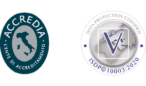
The best all-in-one software platform for 2D and 3D imaging. iRYS is DATA PROTECTION certified and IHE compliant with DICOM networks.
A state-of-the-art tool equipped with a complete ecosystem of features to view, process and share examinations directly from the dedicated workstation, with the computers of the dental practice and with the iRYS Viewer* application available for iPAD.
- Multi-desktop 2D/3D
- Implant simulation
- Compatibility with third parties’ software
- Sharing with 2D and 3D image viewer
- iRYS Viewer for iPad
iRYS is DATA PROTECTION certified and IHE compliant with DICOM networks.
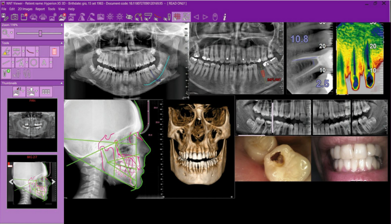
MULTI-DESKTOP 3D/2D
One software to handle 2D and 3D images. The Multi-Desktop system allows for rapid browsing the different 2D and 3D views,
with realistic rendering and multiplanar panoramic analysis. Everything you need to carry out high quality diagnoses and
communicate quickly with the patient.
PRELOADED IMPLANT LIBRARIES
iRYS facilitates the selection and positioning of implants chosen among those contained in its extended library. It is also possible to change them or add new ones in just a few simple steps.
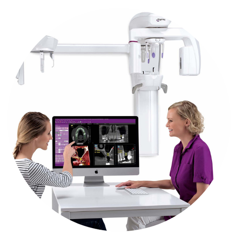
A complete set of tool for your diagnoses.
Simple and efficient diagnosis and planning thanks to the best protocols and the iRYS software filters.
- Filtri immagine evoluti
- PiE (Panoramic image Enhancer)
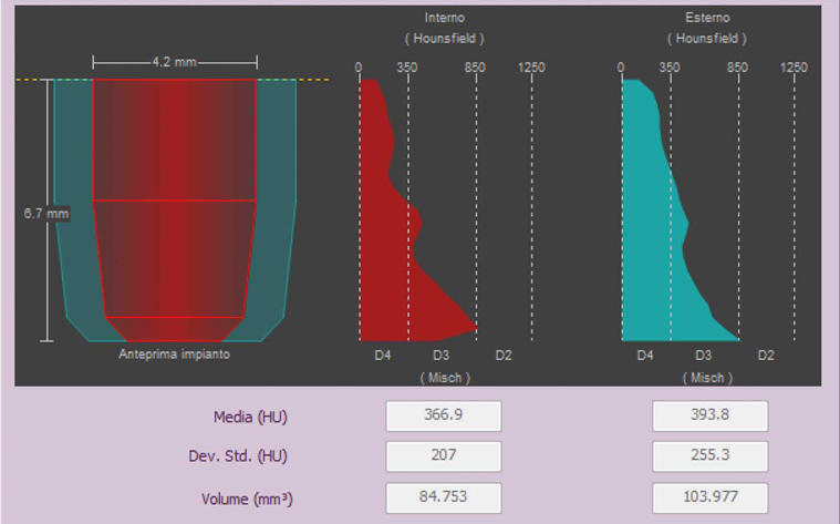
- Bone quality assessment
- Airway volume evaluation
3D SMART
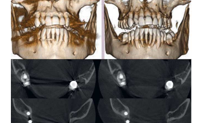
The intelligent 3D SMART (Streak Metal Artifacts Reduction Technology) feature reduces the presence of metal-caused artifacts in 3D volumes through a completely automatic procedure. Make your volumetric images usable at all times, also in the presence of implants and amalgam restorations.
2D PiE
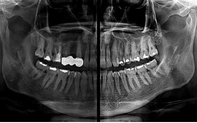
The advanced 2D PiE (Panoramic Image Enhancer) filters allow to maximise 2D image rendering by automatically and selectively optimising the display of different anatomical regions and by making every acquisition detail clearer, from multiple panoramic images to dentition.

AIRWAY VOLUME
iRYS allows to evaluate the upper airways volume in order to investigate possible disorders in the ENT district. This feature is also particularly useful to plan sinus lift surgery in the event of zygomatic implants or for the preliminary assessment of obstructive sleep apnea (OSA).
Case studies
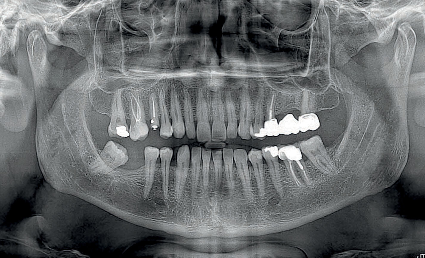
Dental panoramic radiographs
Orthogonal panoramic X-ray: minimises the overlapping of adjacent teeth and provides better periodontal analysis.
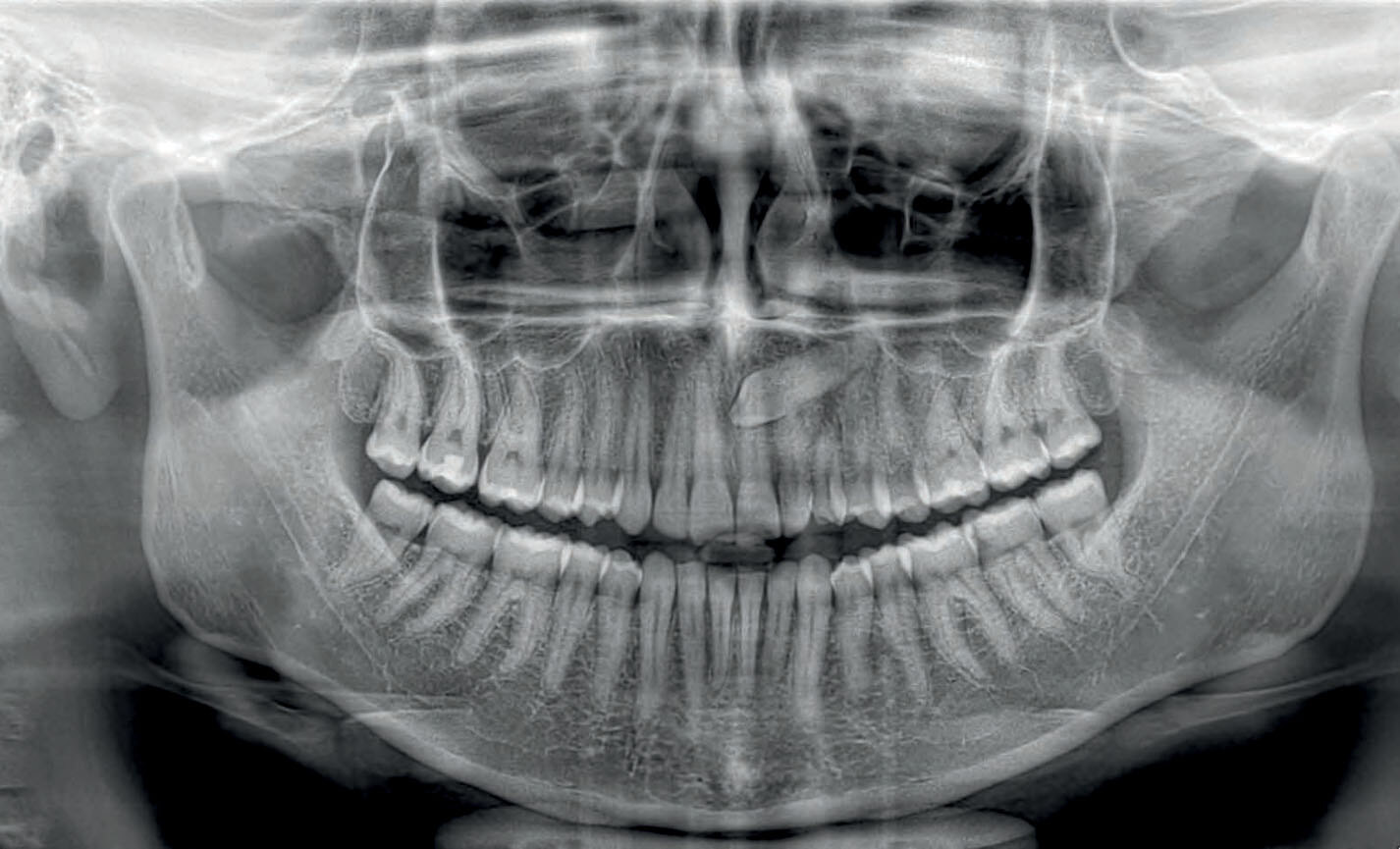
Dental panoramic radiographs
Fast panoramic X-ray: low dose and reduced scan time, perfect for primary investigations, follow-ups or uncooperative patients.
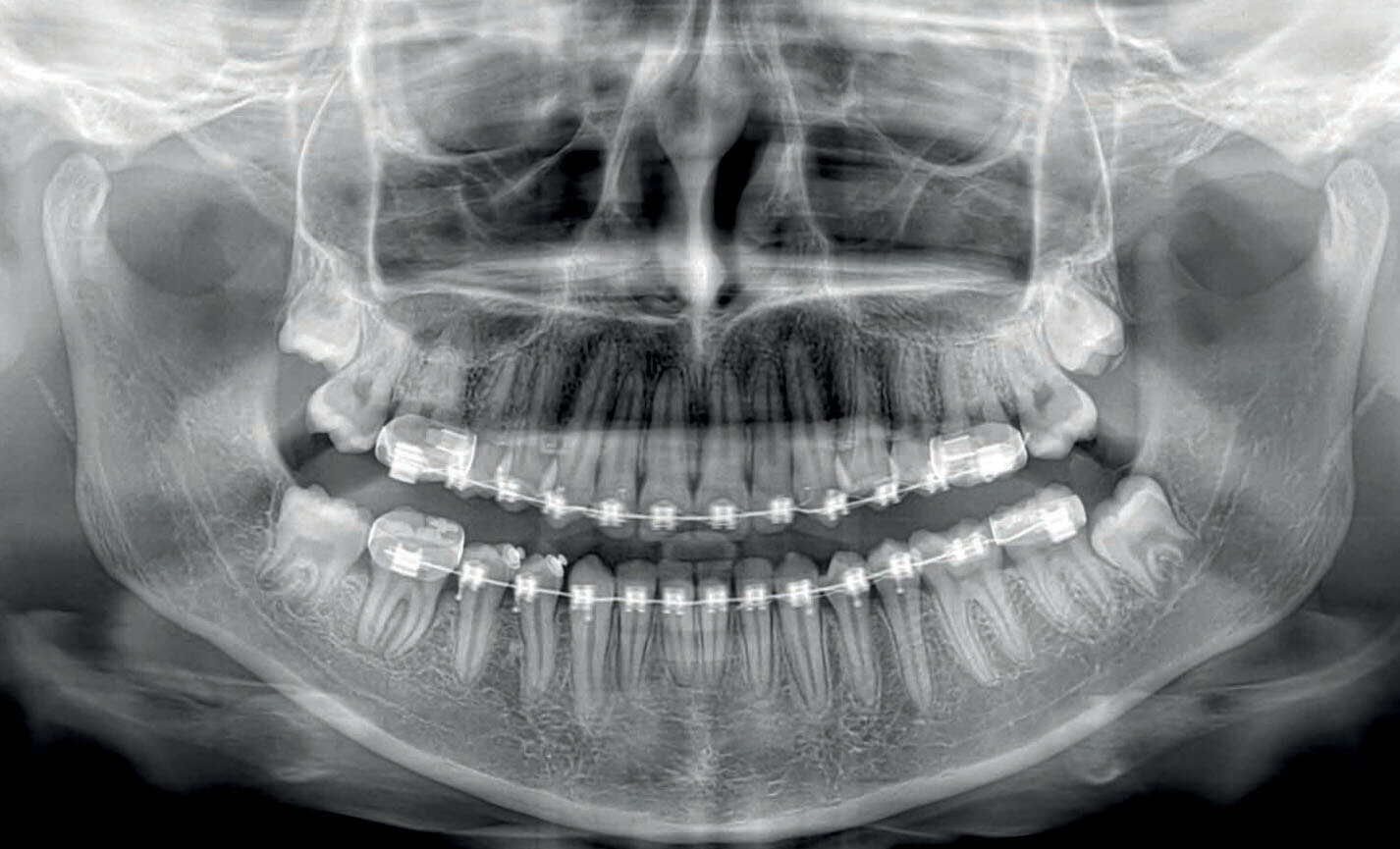
Dental panoramic radiographs
Child panoramic X-ray: limited exposure and optimised parameters for fast paediatric examinations.
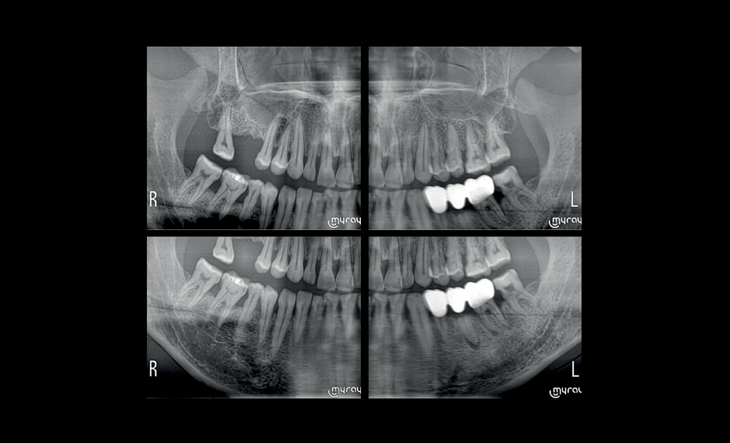
Dental panoramic radiographs
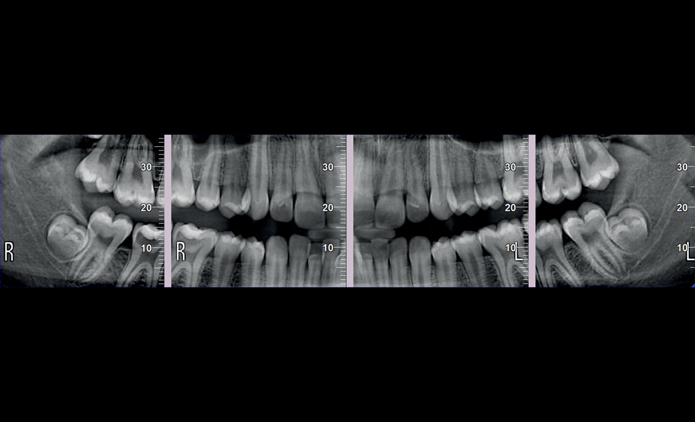
Complete dentition divided into quadrants: localised investigations with selectable segmentation to limit the irradiated dose.
Dental panoramic radiographs
Bitewing projections limited to crowns: high resolution and low dose, a comfortable alternative to intraoral imaging, appreciated by patients with a strong gag reflex.
.jpg?width=1426&height=865&name=01%20(1).jpg)
Stratigrafie extraorali
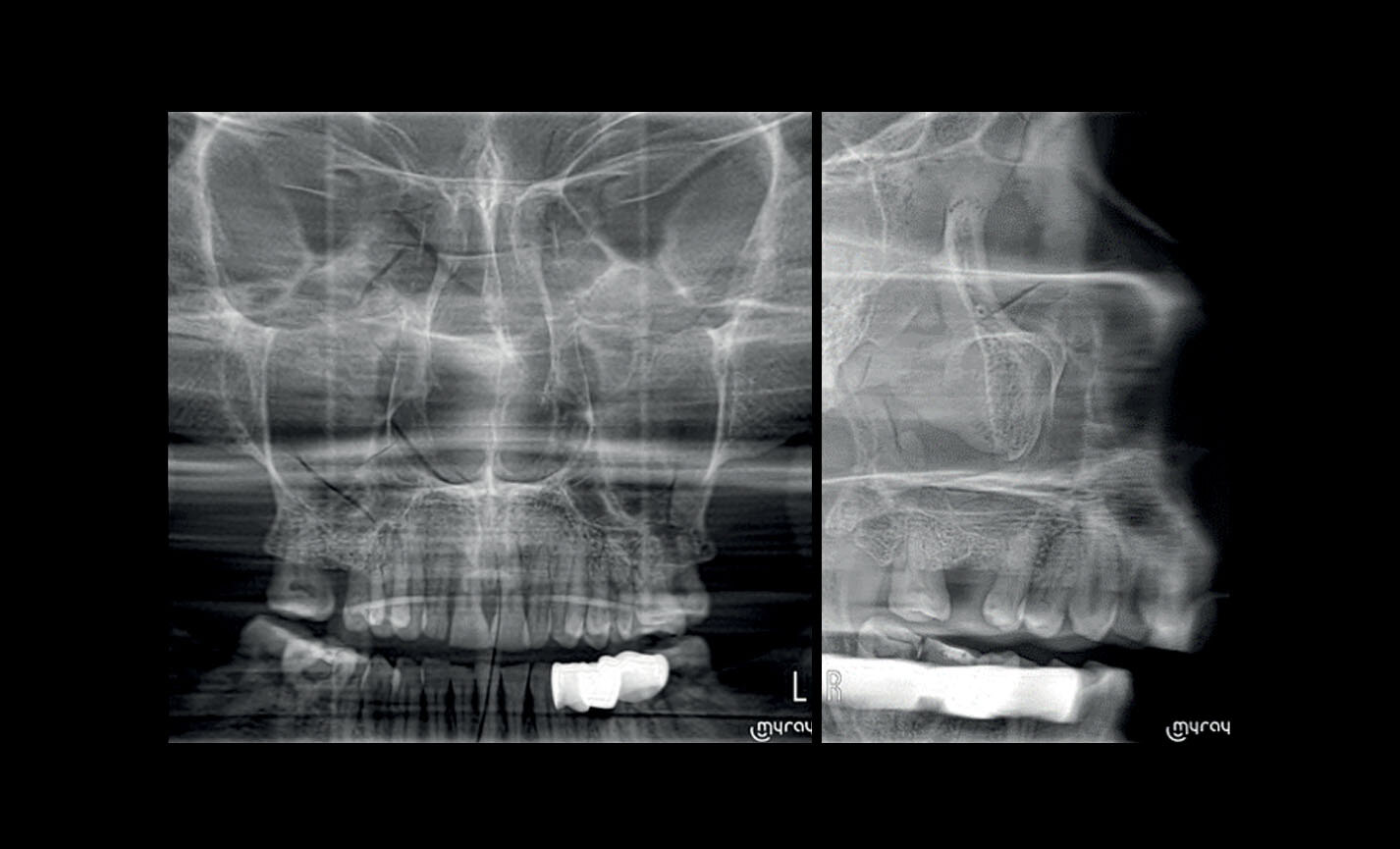
Articolazioni Temporo-Mandibolari: destra e sinistra, a bocca aperta o chiusa ed in proiezione latero-laterale e postero- anteriore con proiezione multi-angolare.
Stratigrafie extraorali
Seni Mascellari: in vista frontale, laterale destra e sinistra, con traiettoria ottimizzata.
Teleradiografia
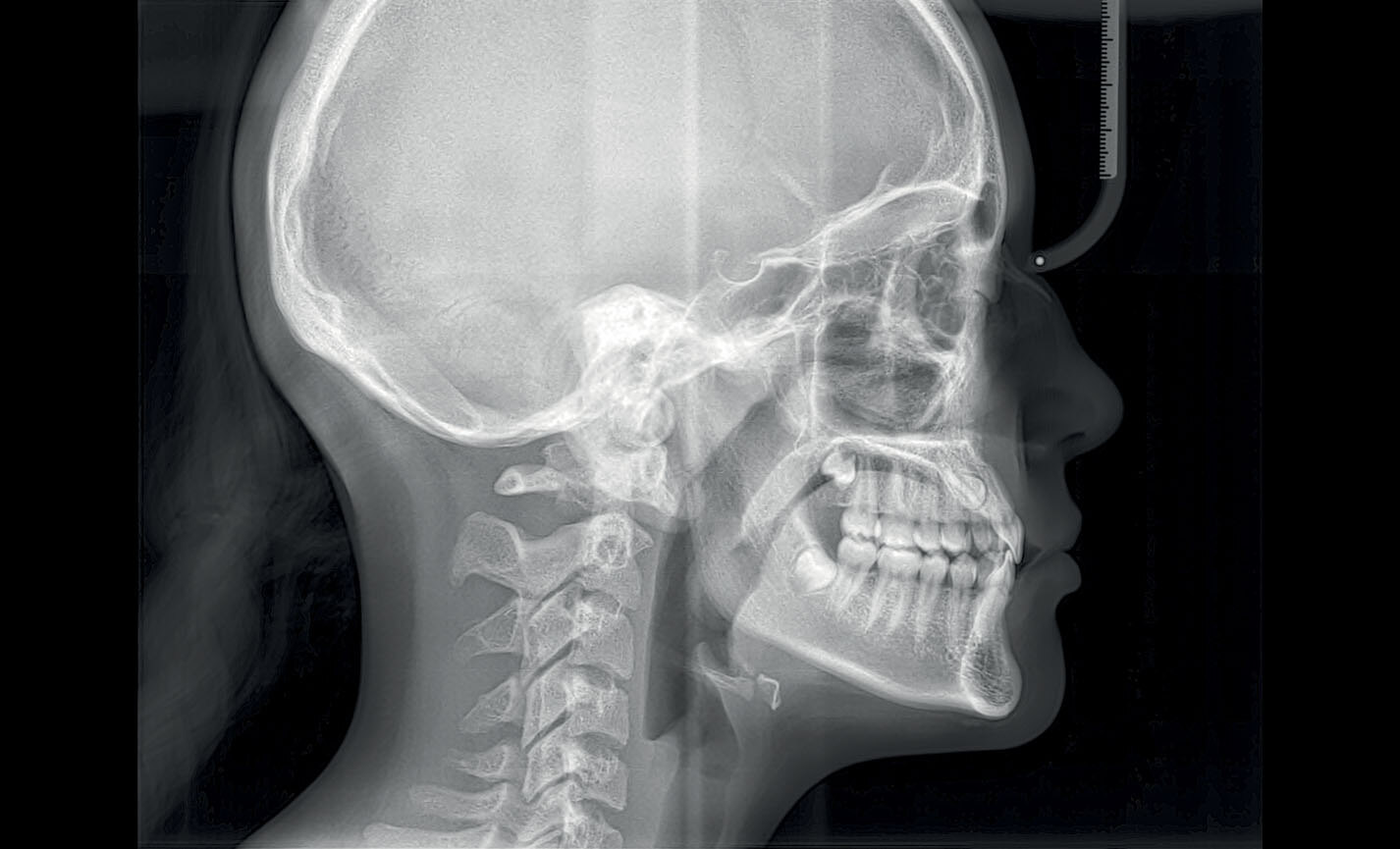
Latero-Laterale: con dettagli ossei e tessuti molli in evidenza, fondamentale per studi cefalometrici.
Teleradiografia
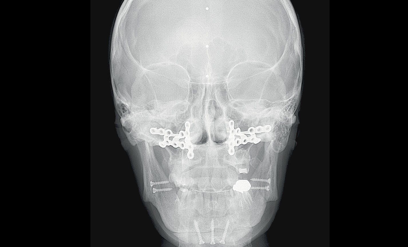
Antero-Posteriore: per indagare asimmetrie e malocclusioni al fine di un corretto trattamento.
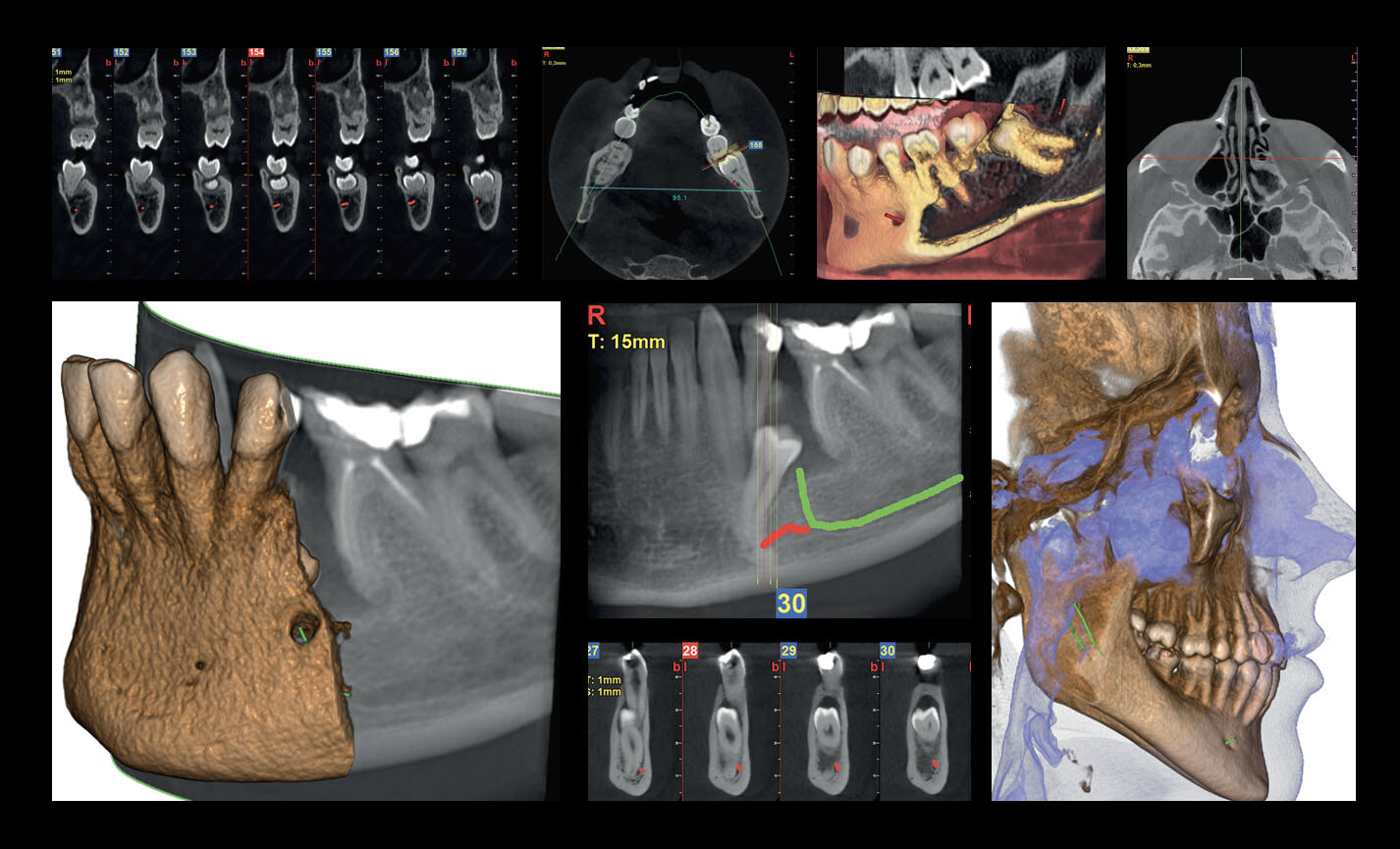
Orthodontic applications
FOVs with a 10 cm diameter are essential for the study of impacted third molars because, in an adult of medium build, the distance between the third molars on the left and right, including the respective roots, the alveolar process and the surrounding bone, is at least 9 cm. Reduced fields of view are useful when analysing impacted or supernumerary teeth in order to restrain the dose to the region of interest. For a correct treatment planning it is indeed crucial to determine the actual position (vestibular or palatal). This is only possible with a 3D analysis, even at a very low dose, with the QuickScan protocol. The complete 13 x 16 cm field of view allows for an accurate assessment of the upper airways, which is often useful to complete the investigation for an orthodontic treatment that does not neglect ENT problems.
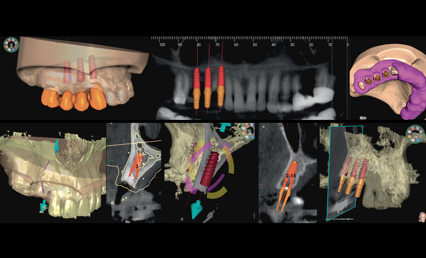
Advanced implant planning
Position the equipment directly on the 3D model, combine it with the STL data from intraoral scanners and define the final prosthetic project. With the advanced implant planning tools you will be able to operate safely thanks to accurate information on the amount of bone and the distance from the surrounding anatomical structures, such as the mandibular canal, defining a minimum safety distance.
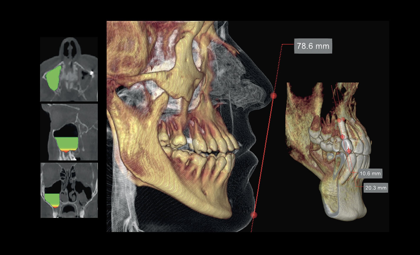
Volume analysis
The software feature for the assessment of the sinus floor lift volume allows for an early planning of the intervention and for a perfectly safe procedure. It is also possible to trace lines directly on the virtual model of the patient thereby assessing morphological relations on the 3D rendering.
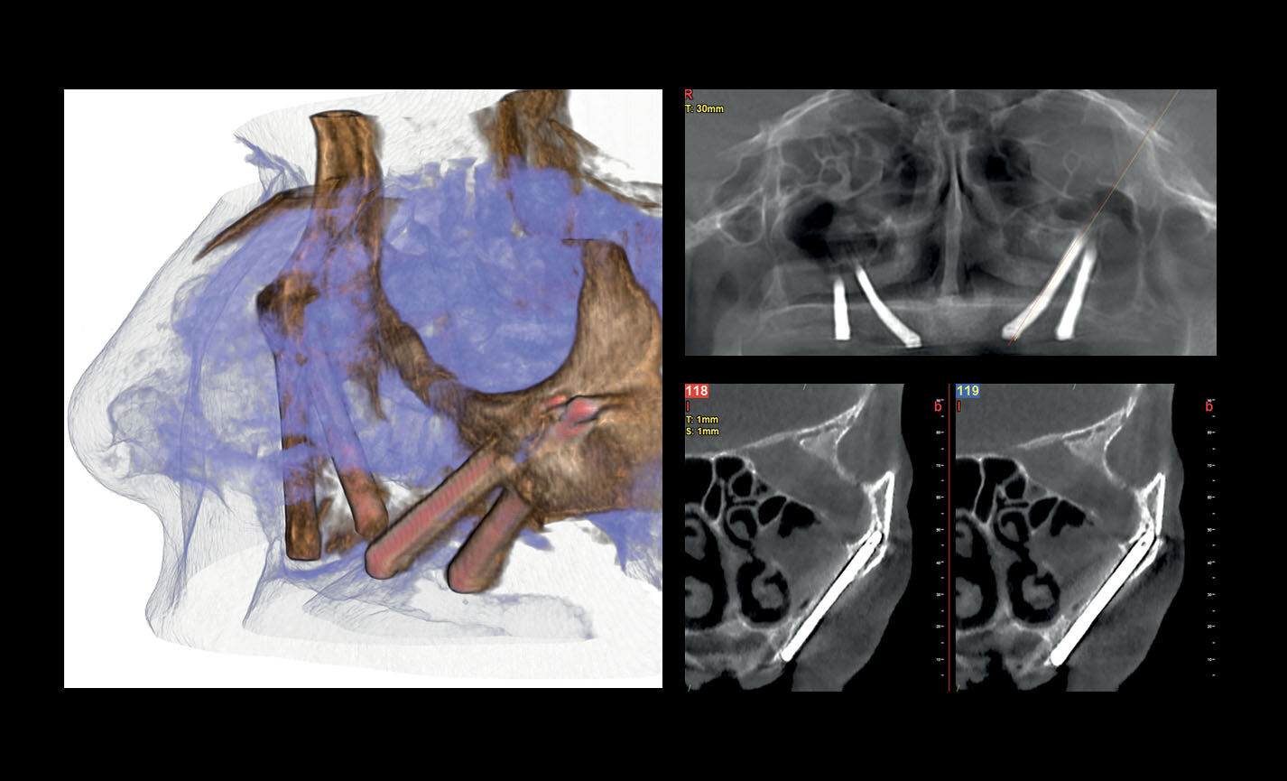
Assessment of zygomatic implants
Volumes with 13 x 8 cm or 13 x 10 cm FOV are the perfect tool for zygomatic implant planning as the 13cm diameter is the only one that makes it possible to include the entire zygomatic arch, without cuts.

