
.png)
Radiographic
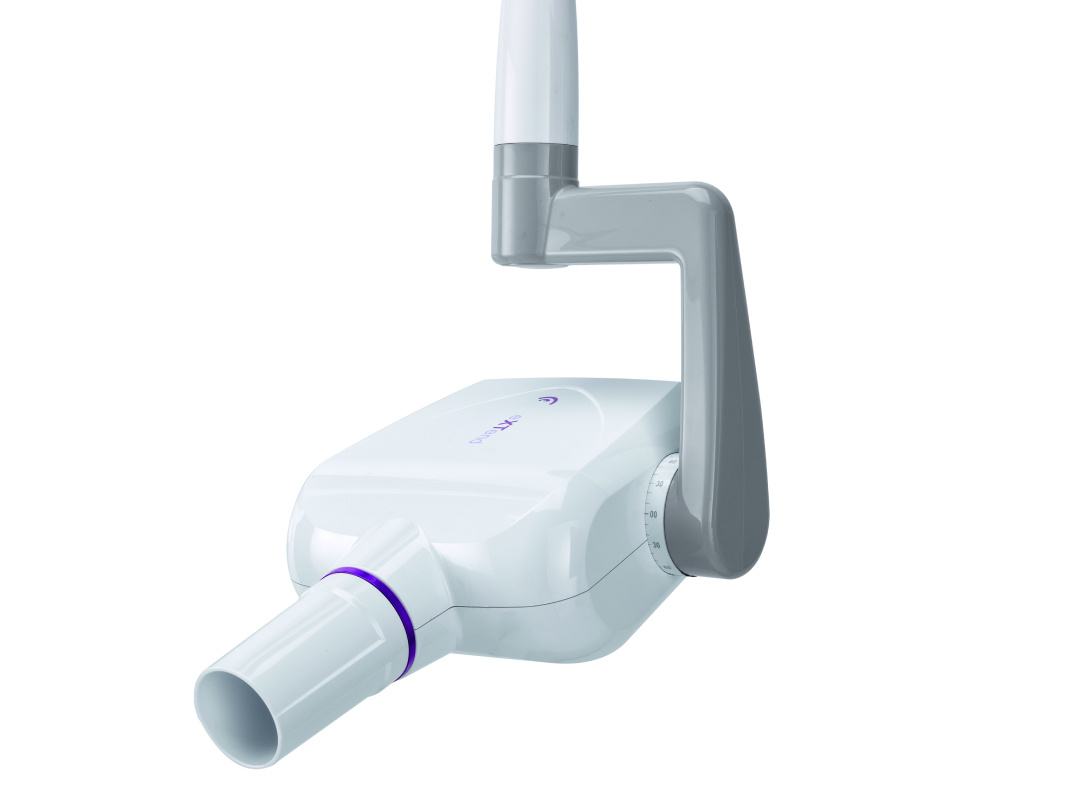
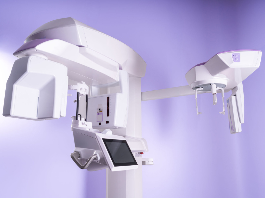
RX DC eXTend
Precise images, total control.
-1.png)

.png)
Precise images, total control.
Automatic modulation
Automatic modulation of exposure parameters adjusts power and timing based on patient size, ensuring sharp images with any sensor.
Less radiation
The high-frequency, constant-potential generator reduces harmful radiation, while the 4 mA mode can be selected to reduce X-ray emission by half. While the rectangular collimator limits the emitted radiation to within the area of interest only, the dynamic duty cycle allows consecutive exposures without overheating, with real-time monitoring of the tube temperature.
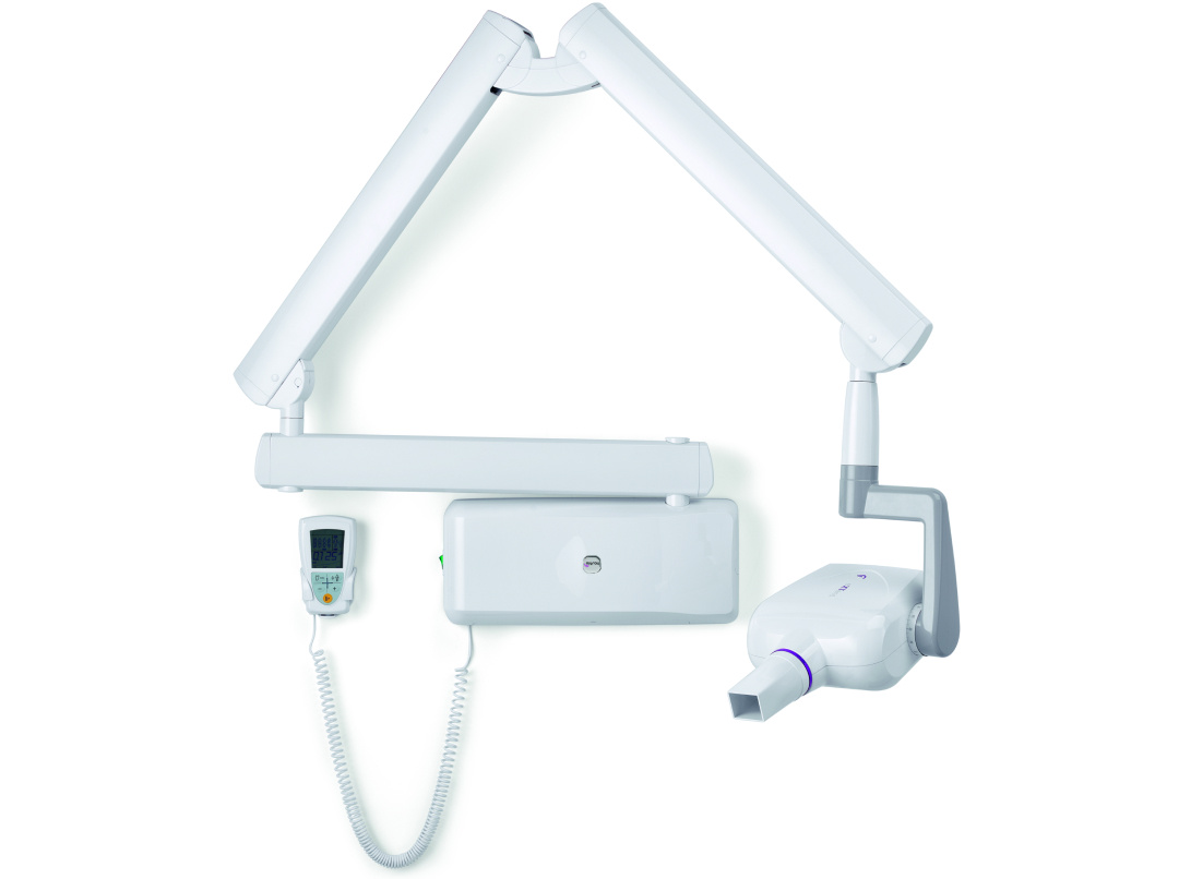
Easy control. Manage your system with the wireless controller, select exposure programs and obtain one-touch instant X-ray images.

Thanks to its clever design and wireless technology, RX DC eXTend is easy to install.
The lightweight, self-balancing arms ensure stability and precision, while the horizontal brackets, available in different lengths (40, 60 and 90 cm), allow seamless integration into any environment.
Thanks to the wall plate suitable for different types of wall installation, RX DC eXTend is designed for easy, uncomplicated setting up anywhere in your surgery.

Built from high-quality materials, RX DC eXTend is designed for strength and durability, minimising vibration and instability during image capturing.
You can count on consistent performance, even under intensive use.

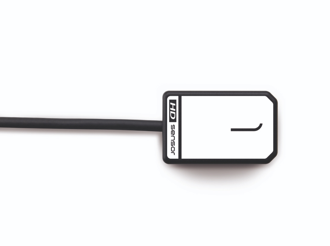
Zen-X DCiS
Zen-X DCiS: the wireless intraoral sensor for precision diagnostics.
Discover the product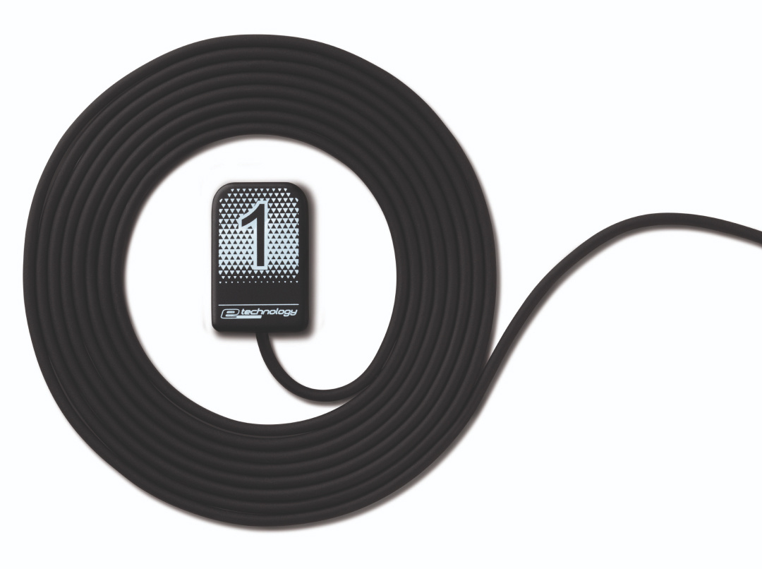
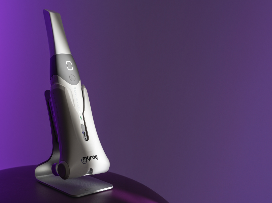
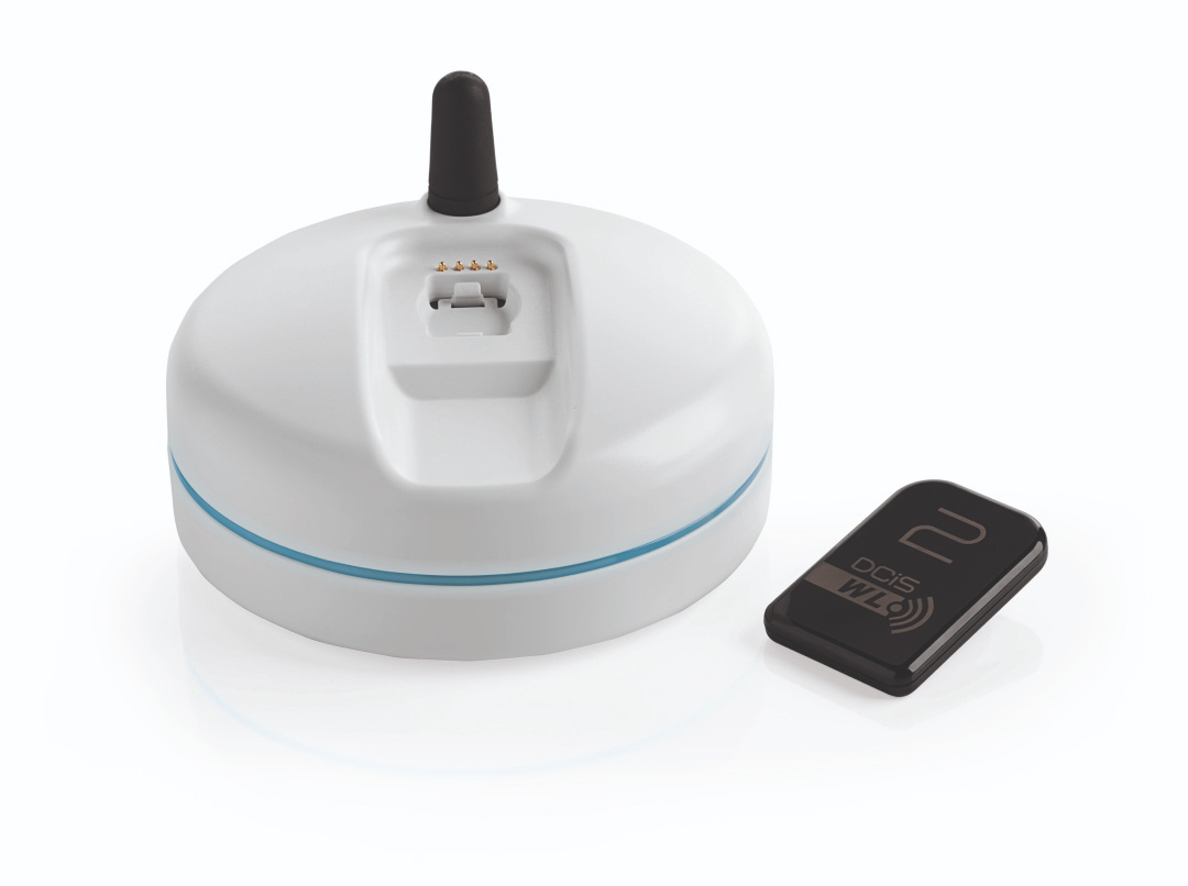
MyScan WL
MyScan WL: wireless digital scanning for a cutting-edge dental clinic.
Discover the product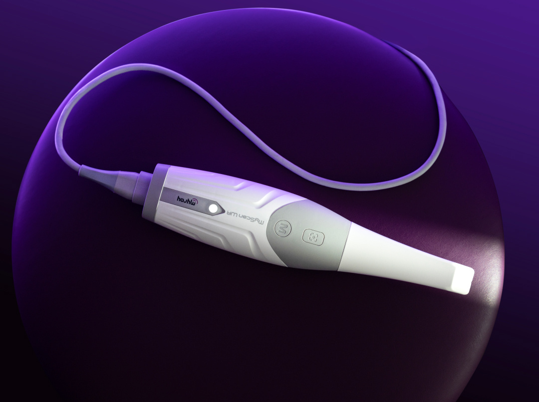
MyScan WR
The power of digital scanning for high-precision diagnostics. With MyScan WR, ergonomics, simplicity and freedom combine to deliver excellent results and an optimal operating experience.
Discover the product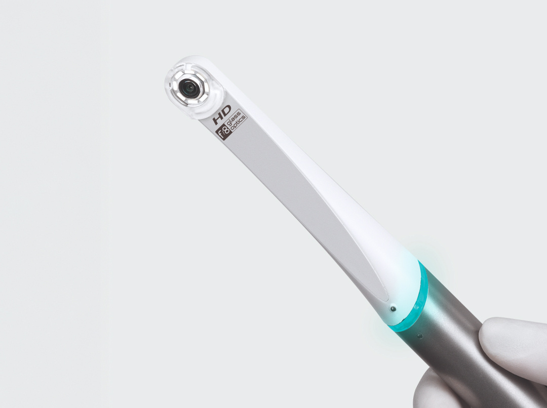

Our dealers are ready to provide you with full support, tailored to your specific needs.